Artificial intelligence increasingly used by doctors as a tool to increase precision in images’ interpretationa
By Deborah Jeanne Sergeant
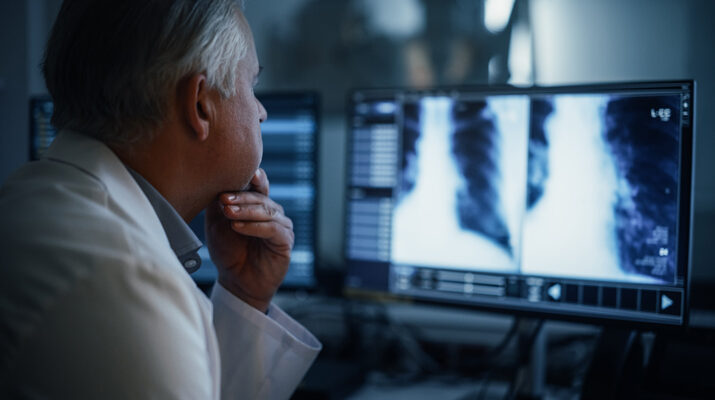
Artificial intelligence has affected many workplaces in a variety of ways. In medical imaging, it has become an integral part of the workday.
“It has been very helpful,” said physician Raymond Tan, chief of the medical imaging department at Highland Hospital. “Right now, we run it in modules. They’re driven for specific paths.”
He said that AI is used for identifying anomalies but not diagnosing problems. That’s up to humans. For example, one module can look for bleeding in the brain, but not explain why a patient is bleeding there.
“These are simpler algorithms than people think,” Tan said. “It makes decision on a higher value of pixels on the scan than normal tissue. It has to locate these things in the correct areas.”
In the past, treatment was given in chronological order of imaging, not in order of medical urgency for treatment reliant upon imaging. AI can help providers know which patients need care faster. For example, someone with a head injury but no bleed would not be prioritized over a patient with a head injury and bleeding.
“Without AI, there was no way to tell who had a bleed and who did not,” Tan said. “Now AI can identify where it suspects there’s a bleed and flag that and move it to the top of our worklist. It’s impactful for the safety of our patients.”
The same applies to blood clots and other emergency symptoms that rely on imaging.
AI can also help providers find potential nodules in the lungs of smokers and ex-smokers. These can be mere millimeters in size.
Tan added that AI can find fractures in the spine and ribs very well and as with the other examples, this can help quickly prioritize patients who are more seriously injured than others in an accident.
Tan reiterated that AI is “not supplanting what radiologist normally do, but it’s more of a helpful aid.”
Although some people fear that AI will eliminate or at least greatly reduce the need for radiologists, Tan disputes this assertion because he believes that the demand for imaging will correlate with the efficiency of reading images.
Jane Bennett, physician, president and CEO of Borg & Ide, a radiology practice in Rochester, said that AI helps read mammography images with a higher level of accuracy for detecting breast cancer.
“We’ve been using now for about three years and I am very comfortable with it,” Bennett said. “I think it makes me a better mammographer. It’s like a second set of eyes on each case. It’s good for us and good for the patient.”
It’s also used for prostate MRI for patients with elevated prostate-specific antigen (PSA). Bennett said these patients used to go “straight to biopsy” but now can receive MRI to get a good look at the lesion.
“If the MRI is negative, they may forgo the biopsy and do sequential MRI to see if there are any changes,” Bennett said. “If it’s suspicious, they can have more targeted biopsy. It’s a less invasive procedure.”
Borg & Ide also use AI for lung cancer screening and cardiac CTA for patients with low to moderate risk.
“It looks at stenosis in the coronaries and assesses whether they’re significant by looking at the physiology of the lesions, drop off and the flow to the stenosis,” Bennett said. “We offer the functional assessment of the coronary arteries and plaque analysis, a newer tool. Some people get calcium scores, but that shows only calcified plaque, not non-calcified plaque. We offer plaque analysis which looks at both.”
She believes that in the next few years, AI will be a tool used in all types of imaging, but never without a radiologist’s assessment and diagnosis.